Buzz! Slap! Ow! Taking the virus out of a mosquito’s bite
MU researchers are getting a deeper understanding how mosquito-borne viruses infect the mosquito - an important step toward eliminating the transmission of mosquito-borne diseases
December 11th, 2018
This video is available for broadcast-quality download an re-use. Closed caption video is also available. For more information, contact Cailin Riley, RileyCi@missouri.edu
COLUMBIA, Mo. – They approach with the telltale sign – a high-pitched whine. It’s a warning that you are a mosquito’s next meal. But that mosquito might carry a virus, and now the virus is in you. Now, with the help of state-of-the-art technology, researchers at the University of Missouri can see how a virus moves within a mosquito’s body, which could lead to the prevention of mosquitoes transmitting diseases.
“Previously, the common understanding was that when a mosquito has picked up a virus, it first needs some time to build up inside the midgut, or stomach, before infecting other tissues in the mosquito,” said Alexander Franz, an assistant professor in the Department of Veterinary Pathobiology in the MU College of Veterinary Medicine and the study’s corresponding author. “However, our observations show that this process occurs at a much faster pace; in fact, there is only a narrow window of 32 to 48 hours between the initial infection and the virus leaving the mosquito’s stomach. For this field of research, that revelation is eye opening.”
What’s Bugging You? from Mizzou News on Vimeo.
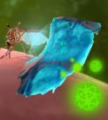
In this study, Franz and the team of researchers observed a mosquito infected with the chikungunya virus, which originates in Africa and was first found in the Americas in 2013. There is no vaccine to prevent or treat this virus, and while most common symptoms include fever and joint pain, they can be severe and disabling. The researchers used three separate electron microscopes to view the virus traveling through the mosquito, beginning with its midgut, or stomach. The first two microscopes provided different two-dimensional views of a single layer of tissue in the mosquito’s stomach. The third, a focused ion beam electron microscope, allowed researchers to see multiple layers of tissue.
“We’re now visualizing a real virus with a three-dimensional model, at scale,” said DeAna Grant, a researcher with the Electron Microscopy Core Facility at the University of Missouri and a co-author on the study. “We can take a three-dimensional image showing the inside of a mosquito’s stomach and say that this dot is a virus particle; there is no guessing to what that dot is. In addition, with this technology we were able to track, in three-dimension, the virus traveling through the mosquito at 24, 32 and 48 hour intervals, and within 48 hours or less, we could see the virus particles leaving the mosquito’s midgut.”
Researchers hope to one day inhibit the genes involved with the release of the virus from within the mosquito’s stomach to prevent future transmission of mosquito-borne diseases. This study is the first life sciences work published using the focused ion beam electron microscope that the University of Missouri purchased through the MU Office of Research in 2016.
The study, “Ultrastructural analysis of chikungunya virus dissemination from the midgut of the yellow fever mosquito, Aedes aegypti,” was published in Viruses. Funding was provided by grants from the National Institutes of Health-National Institute of Allergy and Infectious Diseases (R01AI091972 and R01AI134661), and the University of Missouri award for “Excellence in Electron Microscopy”. The content is solely the responsibility of the authors and does not necessarily represent the official views of the funding agencies.
The study is part of the doctoral project of Asher Kantor, a graduate student in Franz’s lab. In addition to Kantor, Grant and Franz, the publication was co-written by Velmurugan Balaraman of the MU Department of Veterinary Pathobiology, along with Tommi White of the MU Department of Biochemistry and Electron Microscopy Core Facility at the University of Missouri.


 Photos (4):